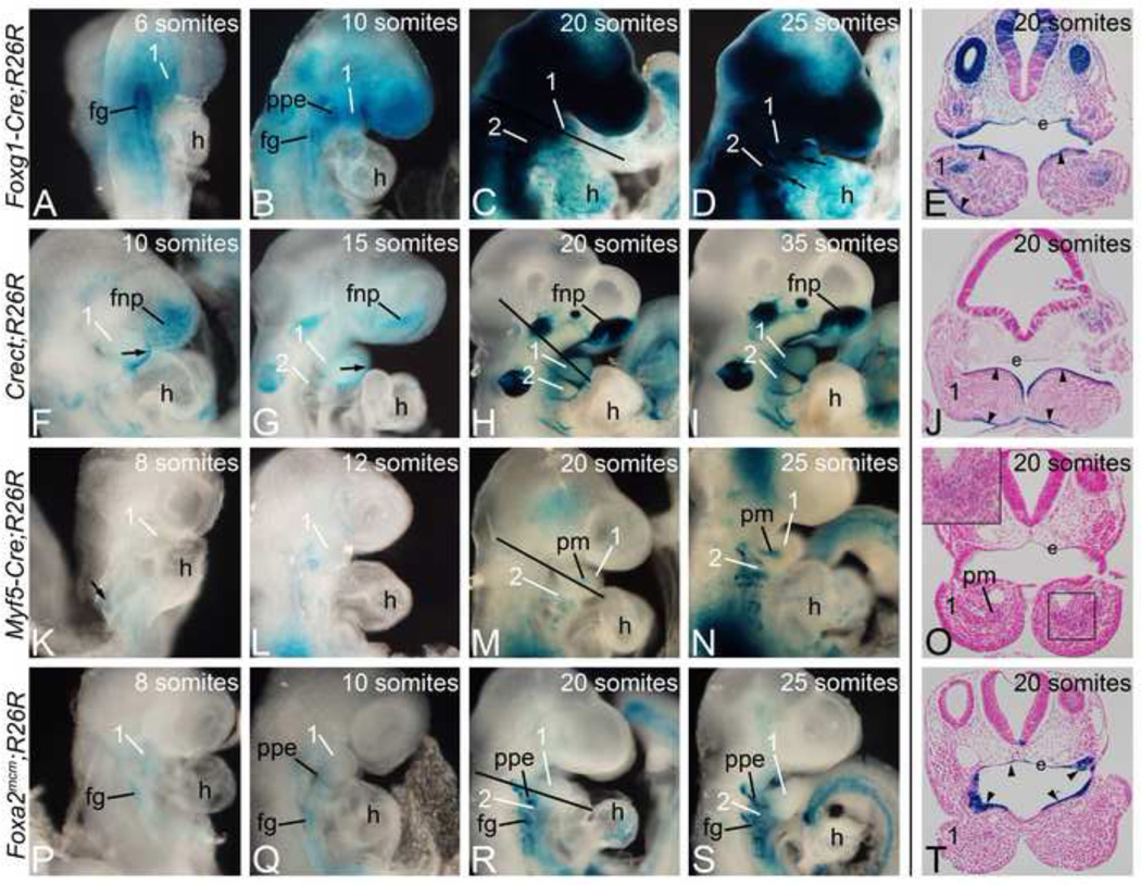
Figure 1. Foxg1-Cre, Crect, Myf5-Cre, and Foxa2mcm activity.
Cre animals were crossed into the R26R Cre reporter strain. Embryos were harvested between E8.0 (6 somites) and E10.5 (35 somites) and stained for β-gal activity. A–D. Cre activity in R26R;Foxg1-Cre embryos was found in endodermal (foregut (fg) and pharyngeal pouch endoderm (ppe)) and ectodermal structures. E. Transverse section through the embryo in C (20 somites) illustrates staining in the arch ectoderm (arrowheads) and endoderm (e), with staining also observed in the paraxial core mesoderm. F–I. β-gal activity in R26R;Crect embryos was found in the ectoderm of the arches (arrow) and in the frontonasal prominence (fnp). J. Transverse section of embryo in H showing arch ectodermal staining at 20 somites (arrowheads). K–N. R26R;Myf5-Cre embryos showed mesodermal staining that at 20 somites could be seen in the paraxial core mesoderm (pm). O. A transverse section through the arches in M illustrated that the staining is restricted to the core mesoderm. (inset). P–S. β-gal activity in Foxa2mcm;R26R embryos starts at E8.0 following tamoxifen injection at E6.5, with endodermal structures labeled (ppe, fg). T. Sections through the first pharyngeal pouch endoderm (e) at 20 somites shows specific β-gal activity (arrowheads). 1, mandibular pharyngeal arch; 2, second pharyngeal arch; h, heart.

