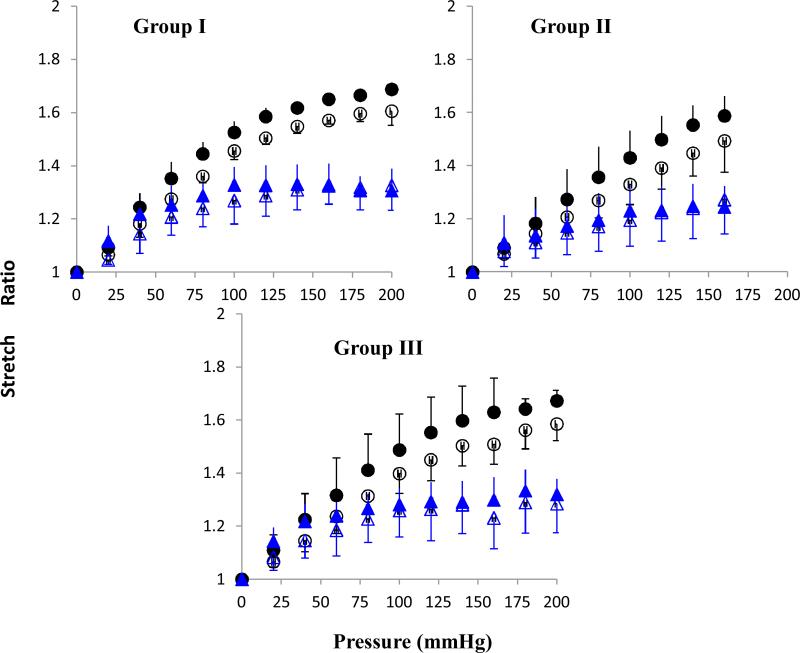
Figure 4.
Axial (circle) and mid-wall circumferential (triangle) stretch ratios plotted as functions of lumen pressure. Hollow and solid symbols represent data pre- and post- treatment, respectively. Three panels shows data (mean ± SD) for arteries in group I (64U/ml, n = 3), group II (128U/ml, n = 4), and group III (400U/ml, n = 6).

