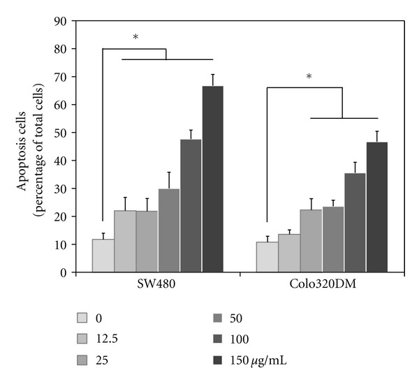
Figure 4.

Detection of apoptotic induction by LCSP. Cells were treated with increasing concentrations ofLCSP as indicated, then incubated at 37°C for 24 h. The treated cells were then suspended and stained with annexin V conjugated with FITC. Ten thousand cells were analyzed by flow cytometry. Data are the averages of three independent experiments and are expressed as the mean ± SD. *Represents a significant difference (P < 0.05).
