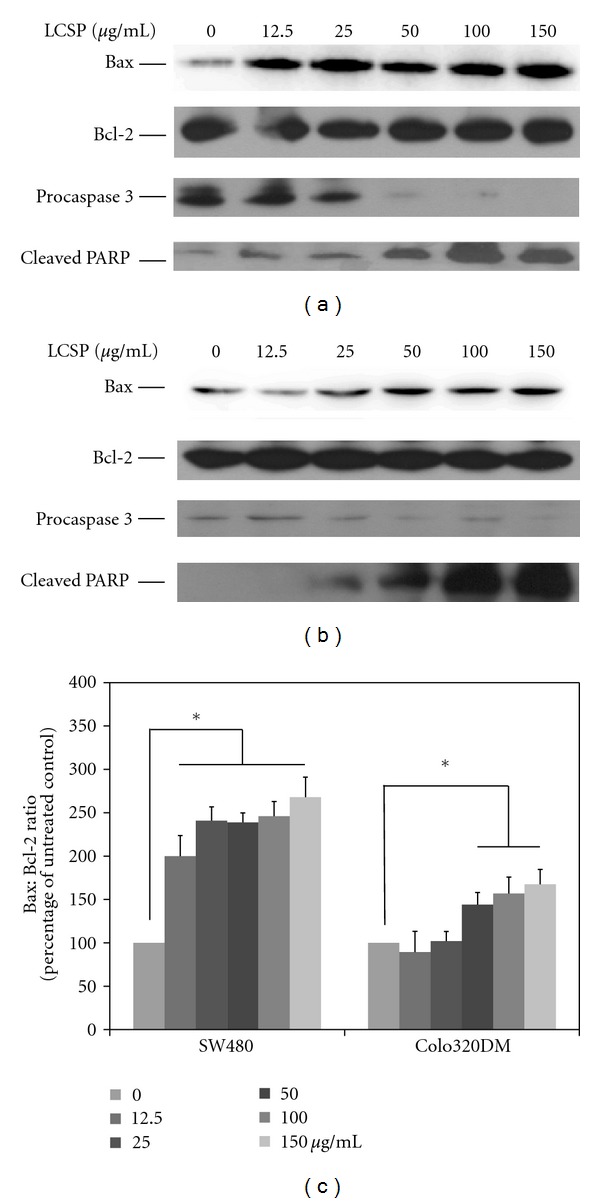
Figure 5.

Immunoblots of apoptosis-controlling proteins in LCSP-treated colorectal carcinoma cells. The cell lysates from LCSP-treated SW480 (a) and Colo320DM (b) were separated by SDS-PAGE, transferred to PVDF membranes, and immunoblotted to show the levels of Bax, Bcl-2, procaspase 3 and cleavage-PARP. The protein levels of Bax and Bcl-2 were quantified using ImageJ software according to the density of each band on the immunoblotting image, normalized to the reference band (β-actin), and presented as the fold of the untreated control. (c) The data reported are the averages of three independent experiments and are expressed as means ± SD. *Represents a significant difference (P < 0.05).
