
Fig. 7.
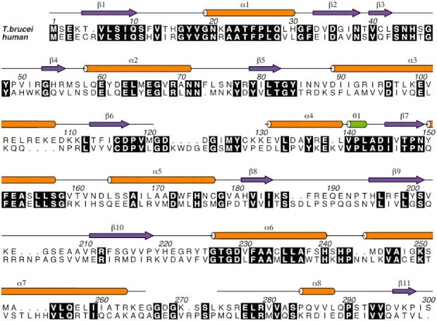
Sequence alignment of TbPdxK with human PdxK. Conserved residues are highlighted in black. Sequences are numbered according to TbPdxK. The secondary structure of TbPdxK is indicated on the top line with α-helices in orange, β-strands in magenta, and 310-helices in green. Figure produced using ALINE (Bond and Schuttelkopf, 2009).