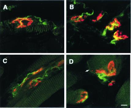
Figure 5.
Laser confocal-scanned images of neuromuscular synapses in tongue muscles of 130-day-old wt (A) and G93A (B–D) mice. Longitudinal sections were incubated with fluorescein-conjugated anti-NF antibody for detection of nerve (green) and rhodamine-α-bungarotoxin (red) to label acetylcholine receptors. (A and B) Typical morphology of normal endplates in a wt mouse (A) and in an age-matched G93A mouse (B). (C and D) Abnormal synaptic configurations in G93A mice. (C) Reinnervation at numerous endplates on a single muscle fiber. (D) An endplate with a thin, unmyelinated terminal sprout is indicated by a white arrow. (Scale bar = 10 μm.)

