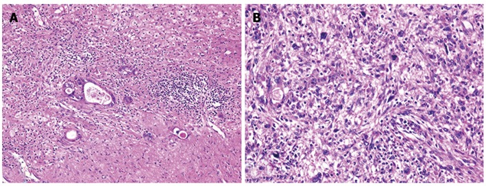
Figure 6.

Histological findings of the tumors and hematoxylin and eosin staining. A: The tumor in segment 8 demonstrated fibrosis and calcification, with a few degenerated residual adenocarcinoma cells; B: The tumor in segment 4 had irregular fascicles of spindle-shaped cells with eosinophilic cytoplasm and nuclear atypia.
