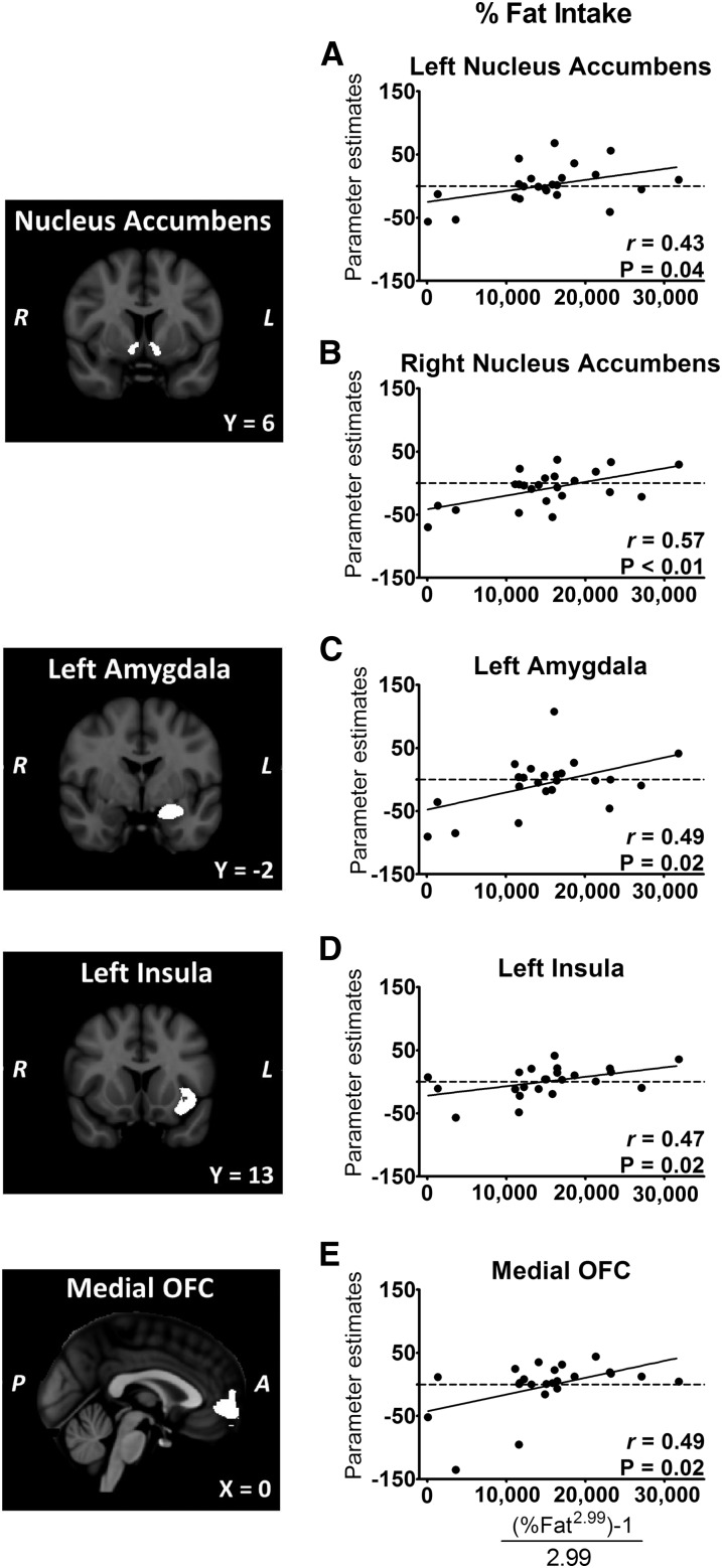
FIGURE 5.
A–E: Greater activation by fattening food cues predicts increased intake of calories from fat. Participants (n = 23) underwent fMRI scans at randomly assigned times after a standardized breakfast, followed by ad libitum intake of foods at a buffet. Left panels: coronal sections of bilateral nucleus accumbens, left amygdala, and left insula ROIs and sagittal section of the medial OFC ROI. Right panels: plots of individual mean ROI parameter estimates for the contrast fattening > nonfattening foods compared with percentage of fat intake. “Fattening” food images depicted high-calorie foods that were previously rated as incompatible with weight loss and considered fattening. Percentage fat intake data (range: 7–46%) were transformed by using a Box-Cox transformation. A, anterior; L, left; OFC, orbital frontal cortex; P, posterior; R, right; ROI, region of interest.

