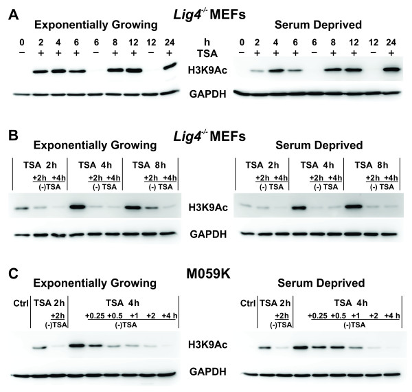
Figure 3 .
Histone H3 acetylation and deacetylation in TSA-treated Lig4-/- MEFs analyzed either during exponential growth (EG) or after serum deprivation (SD).(A) Western blot analysis for histone H3 hyperacetylation (H3K9Ac) after treatment for different times of EG or SD cells with 0.5 μM TSA. Controls (Ctrl) were incubated with DMSO. (B) Time dependent loss of histone H3 acetylation in EG and SD Lig4-/- MEFs after incubation with TSA for the indicated periods of time. Other details are as in 3A. (C) As in panel B but for M059K cells.

