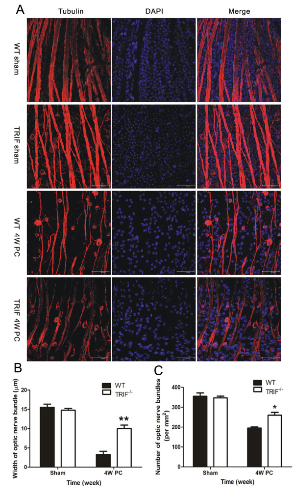
Figure 3.
Optic nerve bundles in whole-mount retina by βIII-tubulin immunostaining. (A) Optic nerve bundles (ONBs) were immunostained with βIII-tubulin and DAPI. In both the WT and trif-/- sham groups, ONBs displayed a regular and full shape. At 4 weeks post-crush (4wPC), both WT and trif-/- ONB became attenuated; this was especially marked in the WT retina, which seems atrophic. Using double-blinded quantification, (B) WT retina were found to display narrower ONBs (B, n = 26) and (C) had a lower value (ρONB; n = 30) compared with those in the trif-/- group.*P < 0.05, **P < 0.01, Scale bar = 50 μm.

