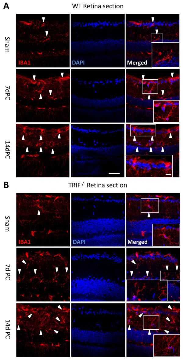
Figure 8.
Microglial cells in retinal sections at post-crush days 7 and 14 (7dPC and 14dPC). Microglia in retinal sections were different between the WT and trif-/- groups at 7dPC and 14dPC. (A) In the WT group, microglia were located in the ganglion cell layer (GCL) and the inner plexiform layer (IPL). After stimulation by ON injury, more microglia were located in the GCL and IPL, and had a ramified and dotted shape at 7dPC. More microglia with a dotted shape and short processes were located in the GCL and IPL at 14dPC. (B) In the trif-/- group, the microglia located in the GCL and IPL had a ramified shape. Scale bar = 20 μm. Scale bar (in box) = 10 μm. GCL, ganglion cell layer; IPL, inner plexiform layer.

