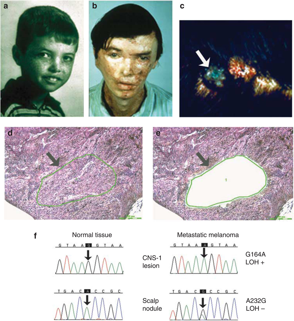
Figure 1. Clinical appearance, histology, and DNA sequencing of metastatic melanoma lesions in patient XP4BE.
(a, b) Comparison of the face of patient XP4BE at the ages of 8 and 27 years. By the age of 27, he had had surgery with grafting for many skin cancers (reproduced from Robbins et al. (1974)). (c) Metastatic melanoma of the scalp before treatment with bis-choroethyl-nitrosourea (arrow). (d) Histology of metastatic melanoma lesion in scalp. Atypical melanocytes are arranged in sheets and nests (arrow) (hematoxylin and eosin (H&E) staining, bar = 200 µm). (e) After capture of the melanoma cells, the remaining tissue was inspected (arrow), and the transfer efficiency of about 500 captured cells was evaluated (H&E staining, bar = 200 µm). (f) Sequencing chromatograms for determination of PTEN mutations. The metastatic melanoma DNA shows a mutation compared with the normal tissue (arrow). LOH, loss of heterozygosity. (Details of method are described in Wang et al. (2009)).

