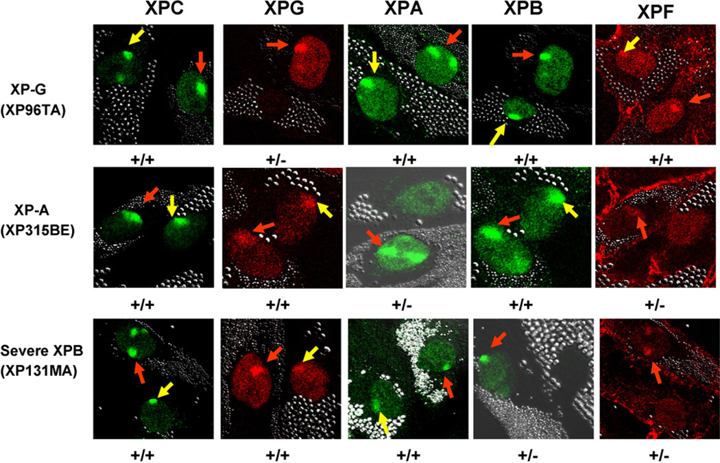
Fig. 5.
Effect of XPG and XPA mutations on recruitment of NER proteins. Normal (0.8 µm beads) and NER deficient XP-G, XP-A or XP-B cells (2.0 µm beads) were treated as in Fig. 2 and fixed immediately (<0.1 h) after UV exposure. Normal cells showed rapid nuclear recruitment of XPC, XPG, XPA, XPB and XPF proteins to the UV damage sites (red arrows). XP-G (XP96TA) cells showed normal recruitment of XPC, XPA, XPB and XPF (yellow arrows) but no staining for XPG protein. In XP-A (XP315BE) cells, normal recruitment of XPC, XPB and XPG was detected (yellow arrows). There was no staining for XPA protein and no recruitment of XPF to the sites of UV damage. In the XP-B cells (XP131MA) from the severe patient, normal recruitment of XPC, XPG and XPA was detected (yellow arrows) (see also Fig. 2B). There was no staining for XPB and no recruitment of XPF protein was detected. (For interpretation of the references to colour in this figure legend, the reader is referred to the web version of the article.)

