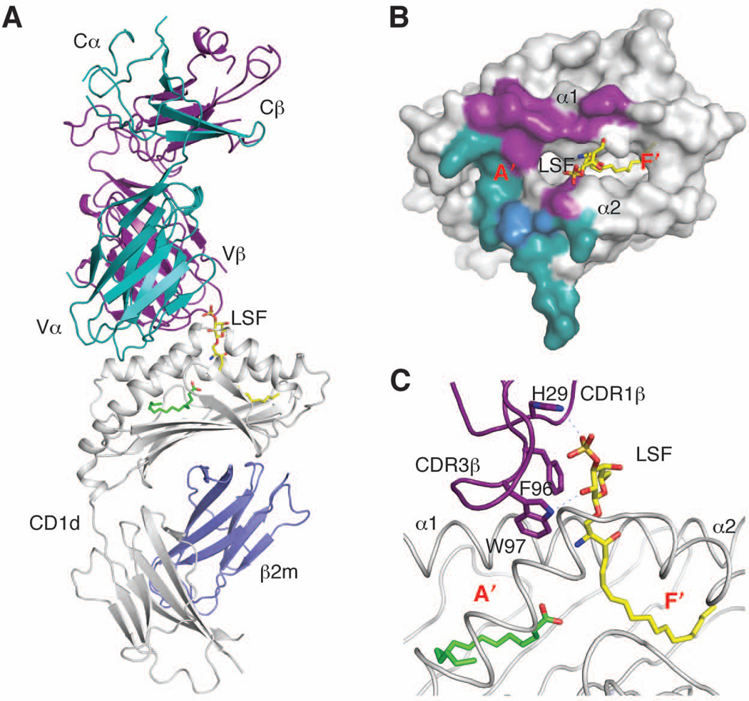
Fig. 3. Antigen recognition by the type II NKT TCR.
(A). Ternary complex (PDB ID 3ELM) between CD1d/b2m (grey and blue), lysosulfatide (yellow), and the type II NKT TCR (α chain in dark cyan, β chain in purple). (B). Footprint of the type II NKT TCR on CD1d. The shared residue Met162 is shown in blue. (C). Detail of the antigen-binding groove showing the bound lysosulfatide in yellow and a spacer in green. The CDR loops and residues contacting the antigen are shown in purple. Polar contacts are shown as dashed lines.

