Figure 6.
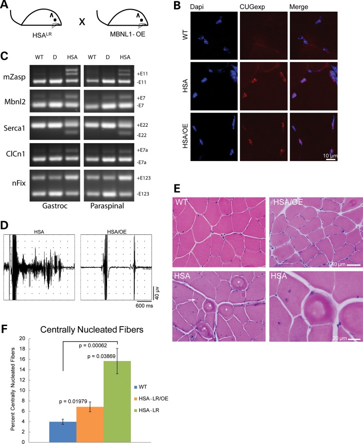
MBNL1 overexpression corrects a DM skeletal muscle phenotype. (A) Schematic diagram of MBNL1-OE and HSALR cross. (B) RNA foci in skeletal muscle from HSALR and HSALR;MBNL1-OE mice detected with a Cy3 conjugated CAG8 oligonucleotide probe. No foci were detected in wild-type FVB mice. (C) RT-PCR splicing assays of FVB wild-type (WT), singly transgenic HSALR (HSA) and doubly transgenic HSALR;MBNL1-OE mice (D). (D) EMG trace from an HSALR gastrocnemius muscle showing a single needle insertion resulting in a ∼2s myotonic discharge and trace from an HSALR;MBNL1-OE doubly transgenic mouse gastrocnemius showing two normal needle insertion responses without electrical myotonia. (E) Hematoxylin and eosin staining of quadriceps muscle from 12-month FVB wild-type, singly transgenic HSALR and doubly transgenic HSALR;MBNL1-OE mice. Ringed fibers (asterisk) and multiple central nuclei (arrow) shown in the HSALR mouse. (F) Quantitation of centrally nucleated fibers in FVB wild-type, HSALR and HSALR;MBNL1-OE mice. Averages are shown ±SEM.

