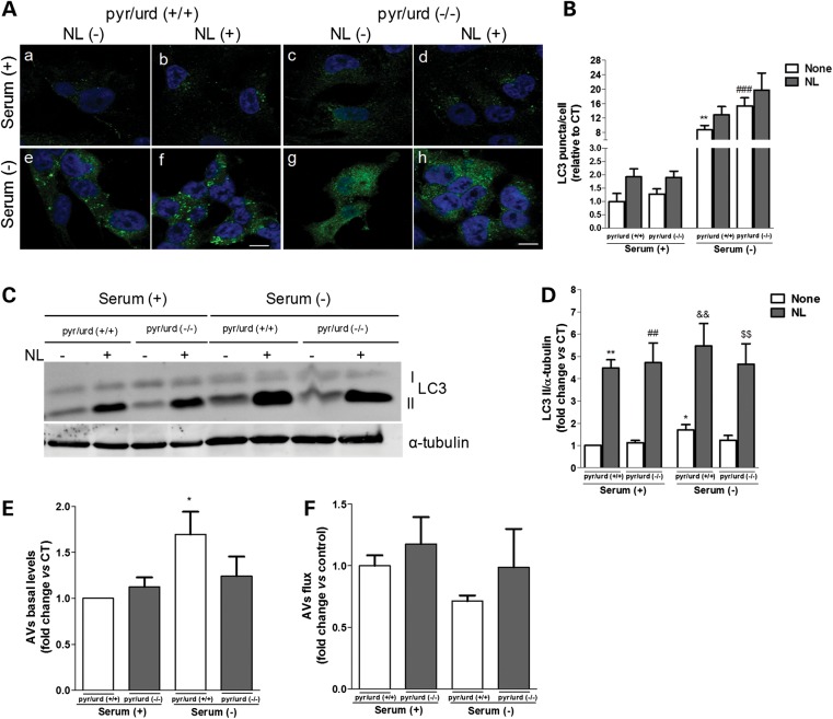
Figure 4.
QC autophagic response is impaired in mtDNA-depleted cells. (A) LC3B immunostaining (green) of Rho0 cells maintained in serum (+) or serum (−) conditions and NL treatment, following growth in the presence [pyr/urd (+/+)] or absence [pyr/urd (−/−)] of pyruvate and uridine. Hoechst 33342-stained nuclei are in blue. (B) Mean number of LC3B-positive vesicles per cell profile [n= 3, **P< 0.01 versus S+ pyr/urd (+/+); ###P< 0.001 versus S− pyr/urd (+/+)]. Scale bar: 10 µm. (C) Immunoblot for endogenous LC3B from Rho0 cells maintained in serum (+) or serum (−) and NL treatment following growth in pyr/urd (+/+) or pyr/urd (−/−) conditions. (D) Densitometric analysis of LC3B endogenous levels [n= 5, *P< 0.05, **P< 0.01, versus S+ pyr/urd (+/+); ##P< 0.01, versus S+ pyr/urd (−/−); &&P< 0.01, versus S− pyr/urd (+/+); $$P< 0.01, versus S− pyr/urd (−/−)]. (E) Determination of autophagic vacuole (AV) levels [n= 5, *P< 0.05, versus S+ pyr/urd (+/+)]. (F) Assessment of autophagic flux (n= 5).

