
Figure 3.
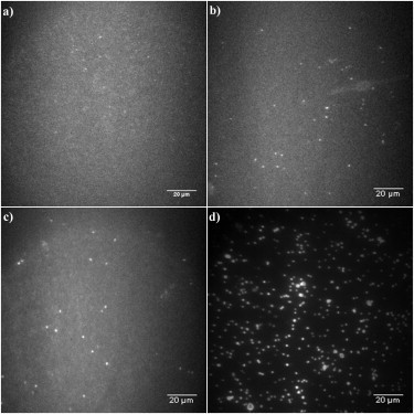
(a) Fluorescence image of the membrane-bound β-amyloid (1–40) peptide, after incubation with 2 nM of β-amyloid (1–40) peptide for 20 h and then washed off, before oligomers formation was observed. (b–d) Fluorescence images showing oligomer formations in the membrane with different concentrations of β-amyloid (1–40) peptide in solution: (b) oligomers formed with 0 nM β-amyloid (1–40) peptides in solution for 130 h; (c) oligomers formed with 2 nM β-amyloid (1–40) peptides in solution for 130 h; (d) oligomers formed with 100 nM β-amyloid (1–40) peptides in solution for 3 h.