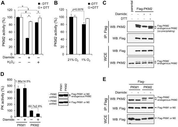
Figure 1. ROS-promoted dissociation of PKM2 subunits and inhibition of enzymatic activity.
(A) A549 human lung cancer cells were treated with 250 μM diamide or 1mM H2O2 for 15 min. After cell lysis pyruvate kinase activity was assayed in the presence or absence of DTT. % PKM2 activity (mean ± SD) relative to untreated cells for each condition is shown (: p<0.001, §: p<0.01, #: p<0.05, 2-way ANOVA with Bonferroni post-tests, N=3).
(B) A549 cells were cultured in medium containing 5.6 mM glucose for 3 hours under 21% O2 or 1% O2. After cell lysis, PKM2 activity was assayed as in (A) (Student’s t-test, N=3).
(C) A549 cells were engineered to express Flag-PKM2 (arrows). Cells were left untreated or were diamide-treated as in (A) and equal portions of lysates prepared under non-reducing conditions were supplemented or not with DTT (1 mM final concentration). Lysates were then subjected to immunoprecipitation with anti-Flag agarose beads in the presence or absence of DTT, followed by SDS-PAGE and western blot.
(D) A549 cells expressing Flag-PKM1 or Flag-PKM2 in the absence of endogenous PKM2 (referred to in the text as Flag-PKM1/kd or Flag-PKM2/kd) were generated as in (17). Expression of Flag-tagged proteins and depletion of endogenous PKM2 were confirmed by western blot with an antibody recognizing both PKM1 and PKM2 (right panel). Left panel: lysates of diamide-treated (250 μM, 15 min.) cells were prepared and assayed for pyruvate kinase activity as in (A). % pyruvate kinase activity (mean ± SD) for each condition relative to untreated Flag-PKM2/kd cells is shown. Numbers denote % difference in PK activity ± SD between diamide-treated versus non-treated Flag-PKM1/kd or Flag-PKM2/kd cells, respectively.
(E) A549 cells expressing Flag-PKM1 or Flag-PKM2 were treated with diamide (250 μM, 15 min.) and, after cell lysis, immunoprecipitation to assess the amount of endogenous PKM2 associated with the tagged PKM isoforms was performed as in (C).

