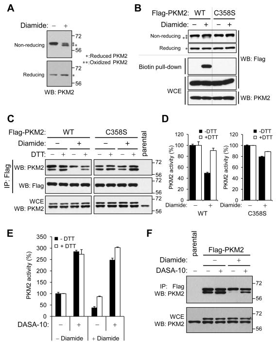
Figure 2. Protection of PKM2 from ROS-induced inhibition by substitution of Cys358 with Ser358 or by small molecule PKM2 activators.
(A) A549 cells were treated with 1mM diamide for 15 min., lysed and analyzed by SDS-PAGE under reducing (lower panel) or non-reducing (upper panel) conditions. Asterisks throughout the figure mark bands corresponding to reduced PKM2 (*) or oxidized (**) PKM2.
(B) Upper panel: A549 cells expressing Flag-PKM2 or Flag-PKM2(C358S) were treated with 250 μM diamide for 15 minutes, lysed, and analyzed by SDS-PAGE under reducing or non-reducing conditions. Lower panel: A549 Flag-PKM2/kd or Flag-PKM2(C358S)/kd cells were treated with diamide as above. Oxidized proteins were labeled with biotin-maleimide (24), purified with streptavidin-sepharose beads and probed for the presence of Flag-PKM2 or Flag-PKM2(C358S) with Flag antibody.
(C) A549 cells expressing Flag-PKM2 or Flag-PKM2(C358S), were treated with 250 μM diamide for 15 min., lysed without DTT and split into equal portions that were supplemented with or without DTT (1 mM final concentration). After immunoprecipitation with anti-Flag agarose, immunoprecipitates were analyzed by reducing SDS-PAGE and western blot with the indicated antibodies.
(D) A549 Flag-PKM2/kd or Flag-PKM2(C358S)/kd cells were treated with diamide as in (C) and pyruvate kinase activity was assayed in cell lysates in the presence or absence of 1 mM DTT. % PKM2 activity (mean ± SD) relative to untreated cells for each condition is shown.
(E) Medium containing 20 μM PKM2 activator DASA-10 (28) was added to A549 cells at t=−1h; diamide was then added directly to the media at a final concentration of 250 μM at t=−15 min. Cells were harvested at t=0, lysed, and pyruvate kinase activity in lysates was assayed in the presence or absence of 1 mM DTT. PKM2 activity is shown as in (D).
(F) A549 cells expressing Flag-PKM2 were treated with DASA-10 and diamide as in (E) and the respective lysates were subjected to immunoprecipitation with anti-Flag agarose under non-reducing conditions. Immunoprecipitates were analyzed by reducing SDS-PAGE followed by western blot with a PKM2 antibody.

