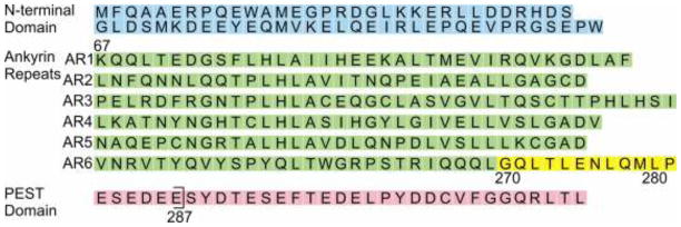
Figure 2.
Amino acid sequence of human IκBα showing the various regions of the protein. The N-terminal domain (not examined in this work) is shown in blue, the ankyrin repeats in green and the C-terminal PEST-like sequence in pink, with the end of the 67–287 construct indicated by a bracket. The residues at the C-terminal end of AR6, for which the analysis was made with CheShift2 (Table 1) are shown in yellow.

