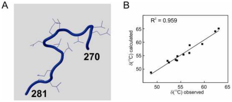
Figure 5.

Structure of the segment Gly270 to Pro281 of IκBα, derived from 1IKN (Huxford et al., 1998) colored by CheShift-2, which discriminates small (blue), medium (white) and large (red) deviations from the structure expected on the basis of the chemical shifts. B. Correlation of the observed chemical shifts (Sue and Dyson, 2009) and those calculated from the 1IKN structure using CheShift-2.
