
Figure 5.
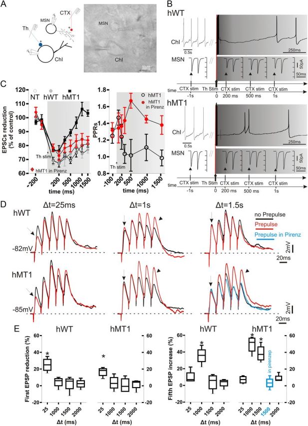
Thalamic stimulation unbalances muscarinic receptor-mediated modulation at corticostriatal synapses. A, Paired recordings were obtained from a ChI and a neighboring MSN during both cortical (CTX, red) and thalamic (Th, blue) stimulation. IR image of the slice preparation (dotted lines mark recording pipettes). B, In hWT mice, the ongoing firing activity of a ChI was interrupted when CTX stimulation was preceded by thalamic stimulation (50 Hz, 10 pulses; gray traces, top right). Such protocol resulted in a decrease of the EPSC amplitude (bottom traces), persisting for up to 1 s after thalamic stimulation (bottom traces). In hMT1 mice (bottom), a similar protocol induced a brief pause in ChI, and a rebound spiking activity. The reduction in EPSC amplitude was smaller than in hWT mice, and declined within 1 s after thalamic stimulation (bottom traces). C, Left, Summary graph of the time dependence of EPSC reduction in the three genotypes, and in hMT1 mice in pirenzepine (red diamonds). C, Right, PPR measurement in hMT1 mice suggests a presynaptic effect at 100 ms, but not at later measurements (500 ms–1.5 s). The presynaptic M2-dependent effect was restored in pirenzepine (red circles). D, A thalamic prepulse (50 Hz, 10 pulses) was given at three times (Δt = 25 ms; Δt = 1 s; Δt = 1.5 s) before the corticostriatal stimulus (5 consecutive pulses, 20 ms interval). Only at short delay (25 ms) a decrease in EPSC amplitude was recorded, suggestive of a presynaptic inhibition in both genotypes (arrows). However, at longer delays (Δt = 1.5) a significant enhancement of EPSP amplitudes was observed only in hMT1 mice (arrowheads). Note that the increase in EPSP amplitude was prevented in the presence of pirenzepine (blue trace). E, Plots summarize the changes (%) in amplitude of the first and fifth EPSP, respectively, at different prepulse delay times.