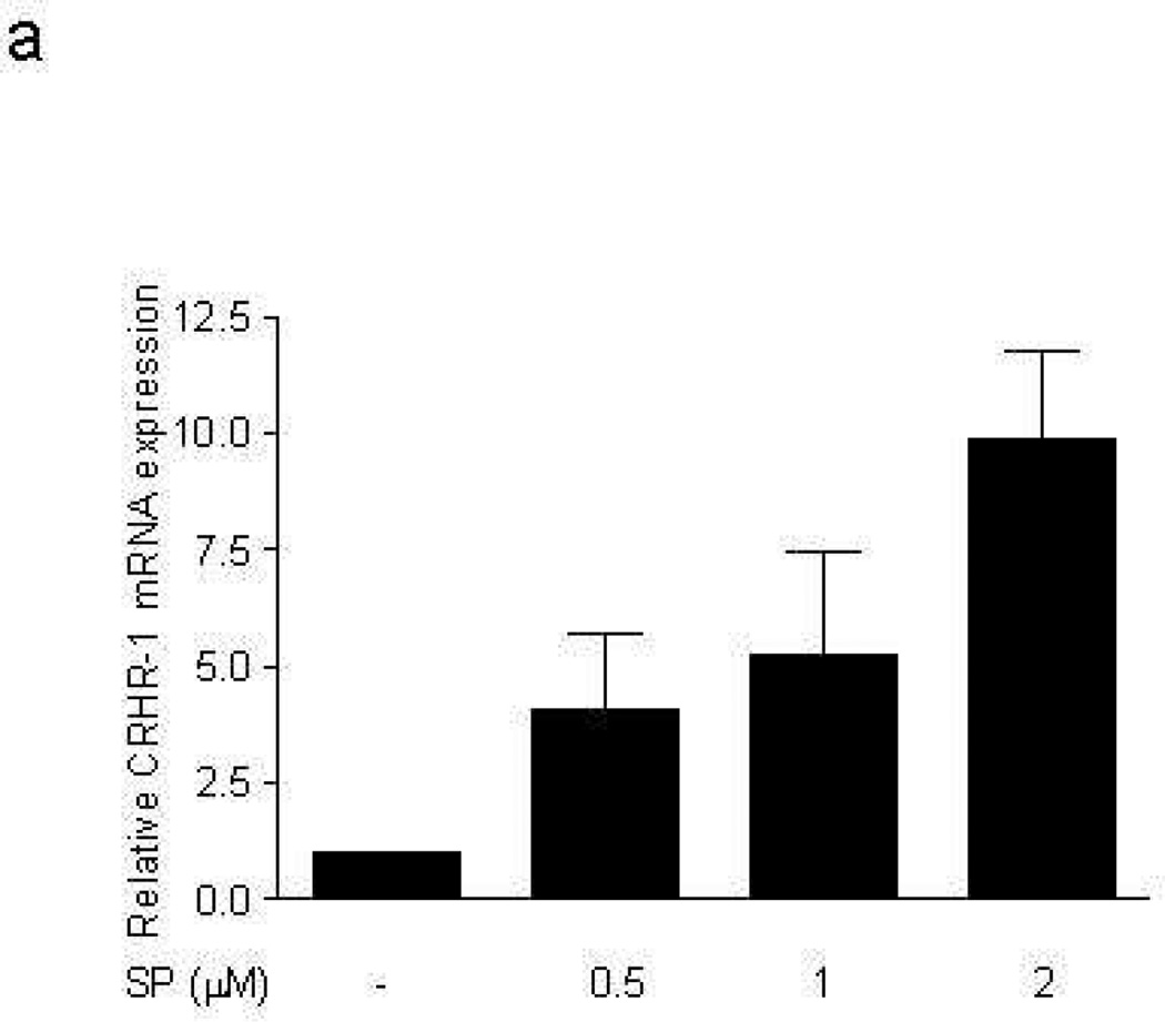
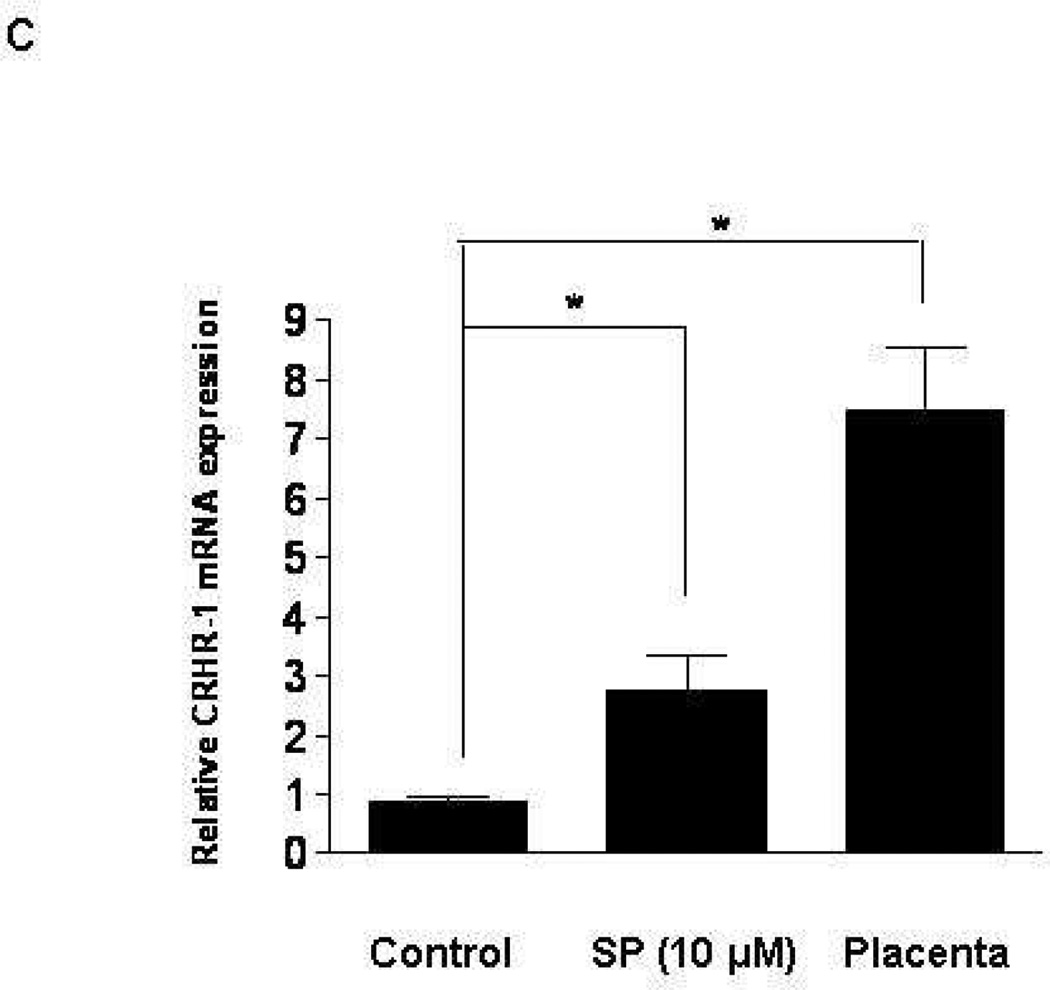
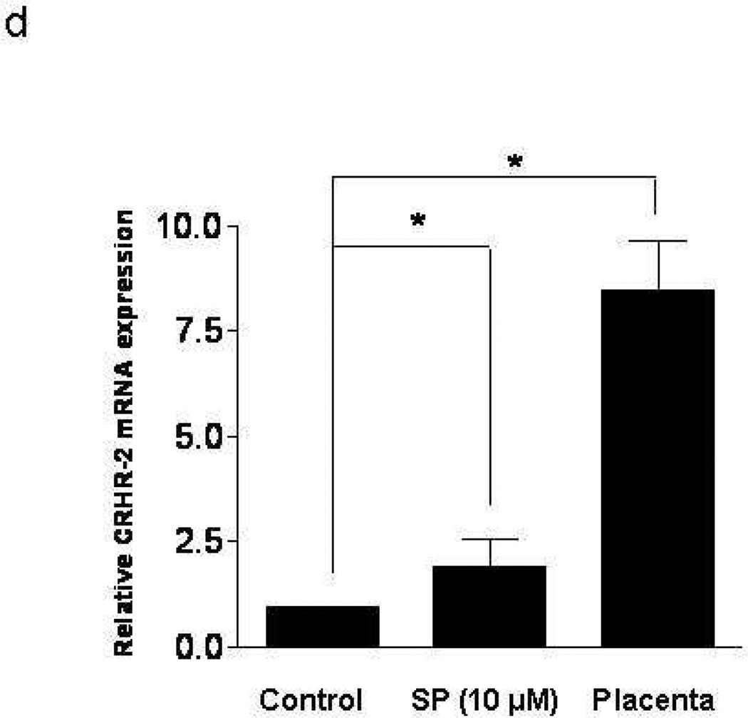
Figure 1. Effect of SP on CRHR-1 mRNA expression in human mast cells.
LAD2 cell expression of CRHR-1 (a) mRNA following treatment with SP (0.5, 1, 2 µM) for 6 hr and (b) protein following treatment with SP (0.5, 1 µM) for 48 hr; y-axis indicates “counts” of cells, while the x-axis indicates log fluorescence intensity (n=3, p<0.05). (c,d) Ten week-old hCBMCs expression of CRHR-1 and CRHR-2 mRNA, before and after 6 hr incubation with SP (10 µM) at 37°C. Human placenta was used as a positive control for CRHR-1 and CRHR-2. Gene expression was analyzed by RT-PCR and expression is indicated relative to control (n=3, p<0.05).


