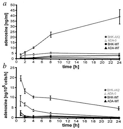
Figure 1.
The accumulation of adenosine in the supernatants of cultured cells. (a) The amount of adenosine in the supernatants of BHK-AK2 (shaded circles), ADA-O (▵), and their WT counterparts, BHK-WT and ADA-WT cells (● and ▴, respectively) was measured over 24 h by using an enzyme-coupled fluorescent assay from 105 cells each. (b) The adenosine levels determined under a were recalculated to represent the amount of adenosine that accumulated within a time interval (ng of adenosine per 105 cells per h). Each release study was performed six times; bars were calculated as ±SEM.

