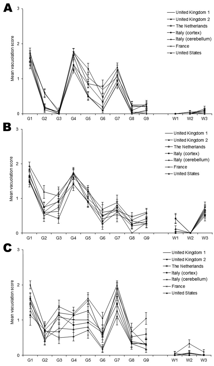
Figure 2.
Lesion profile comparison of variant Creutzfeldt-Jakob disease cases show similarities in vacuolar pathology levels and regional distribution in mouse brains. Wild-type mouse lines RIII (A), C57 (B), and VM (C) are shown. Data show mean lesion profile ± SEM (n>6). G1–G9, gray matter scoring regions: G1, dorsal medulla; G2, cerebellar cortex; G3, superior colliculus; G4, hypothalamus; G5, thalamus; G6, hippocampus; G7, septum; G8, retrosplenial and adjacent motor cortex; G9, cingulate and adjacent motor cortex. W1–W3, white matter scoring regions: W1, cerebellar white matter; W2, mesencephalic tegmentum; W3, pyramidal tract.

