
Fig. 3. CIE disrupts mPFC extinction encoding and decreases mPFC NMDAR-mediated currents.
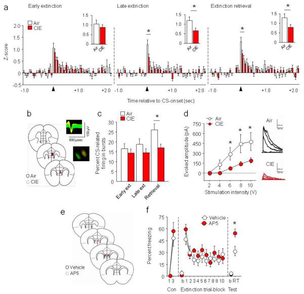
(a) In vivo freely moving recordings of IL neurons demonstrated CIE decreased neuronal activity during the first 100 milliseconds (redrawn in insets) of CS onset during late extinction (trial-block 10) and extinction retrieval (air n=7, CIE n=7, n=69-85 neurons, for a-c). (b) Coronal cartoon showing electrode placements in IL. Example extracellular waveforms and principal components separation of simultaneously recorded neurons. (c) CIE decreased CS-related IL neuronal bursting during extinction retrieval. (d) CIE decreased stimulation-evoked synaptic NMDAR-mediated currents in IL neurons, with example traces (air n=7, CIE n=8). (e) Coronal cartoon showing cannula placements in IL. (f) NMDAR antagonist AP5 infused into IL immediately after extinction training impaired extinction retrieval in alcohol-naive mice (air n=8, CIE n=7). Data are Means ±SEM. *P<.05 CIE versus air