
Fig. 2.
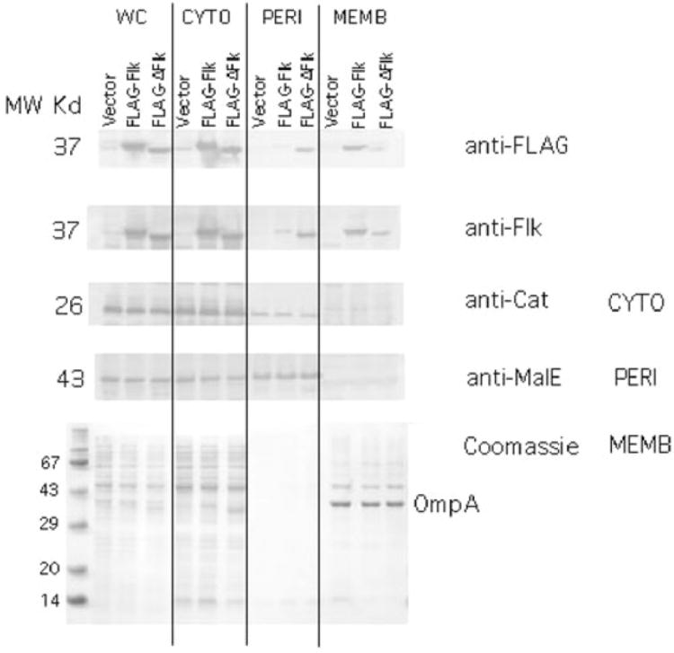
Cellular fractionation of the Flk protein. Plasmids pJK462 and pJK463 express Flk or FLAG–flk from the T7 promoter respectively. These plasmids and a vector control, pJK447 were placed in strain TH4657 that harbours T7 RNA polymerase under control of the lac promoter (Table 2). Following induction of T7 RNA polymerase, the whole cells (WC) were separated into cytoplasmic (CYTO), periplasmic (PERI) and membrane (MEMB) fractions (Experimental procedure). The proteins chloramphenicol acetyl transferase (Cat), maltose binding protein (MalE) and outer membrane protein A (OmpA) were used as controls for proteins known to be cytoplasmic, periplasmic and membrane-associated respectively. The fractions were run on SDS-PAGE and analysed by Western blot with anti-FLAG, anti-Flk, anti-Cat, anti-MalE antibodies. MW, molecular weight.