
Fig. 4.
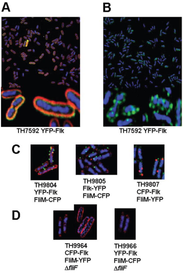
Flk appears as discrete spots in the cell membrane.
A. Expression of YFP–Flk is associated with the membrane fraction as indicated by the membrane stain FM 4-64.
B. Visualization of YFP–Flk in the absence of membrane stain indicates that it forms discrete spots on the cell membrane.
C. Strains TH9804 expressing an N-terminal YFP fusion to Flk (YFP–Flk) and a CFP-labelled HBB (FliM–CFP), TH9805 expressing a C-terminal YFP fusion to Flk (Flk–YFP) and TH9807 expressing an N-terminal CFP fusion to Flk (CFP–Flk) and a YFP-labelled HBB (FliM–YFP).
D. Strains TH9964 and TH9966 express CFP–Flk and FliM–YFP or YFP–Flk and FliM–CFP respectively, but are also deleted for the fliF gene, which is required for FliM assembly into HBB structures. Strain TH9964 was stained either with DAPI and the CFP–Flk visualized in red (TH9964, left photo) or with FM 4-64 and CFP–Flk visualized in blue (TH9964 right photo). TH9966 was stained with DAPI and YFP–FLK visualized in red. For TH9964 or TH9966, FliM–YFP or FliM–CFP, respectively, was not detected.