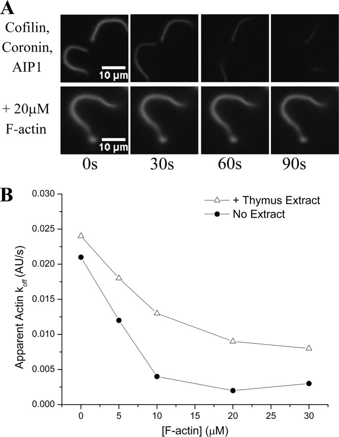
FIGURE 1.
Physiological concentrations of F-actin inhibit disassembly of an F-actin substrate; an unknown factor protects against this inhibition. A, cofilin, coronin, and AIP1 readily disassemble a fluorescently labeled actin substrate (L. monocytogenes comet tails) on the required cellular time scale of tens of seconds (top series) but are inhibited when challenged with a physiological excess of F-actin (bottom series). B, Actin disassembly activity of this same mixture is markedly sensitive to increasing concentrations of F-actin, nearing complete inhibition by 10 μm F-actin (circles). The addition of a high speed supernatant derived from thymus extract, however, protected against this inhibition (triangles). Experiments were done in the presence of cofilin (2 μm), coronin (2 μm), and AIP1 (200 nm) with prepolymerized F-actin at the concentrations indicated. AU, arbitrary units.

