FIGURE 2.
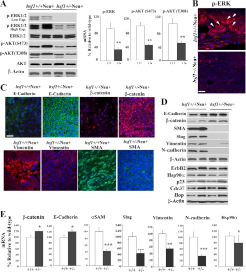
Mammary tumors from Hsf1+/−Neu+ mice exhibit lower ERK1/2 activation and reduced EMT. A, tumor cell extracts from the indicated genotypes were used in representative immunoblotting to detect expression of total and phosphorylated ERK1/2 (low and high exposures (Exp.) are indicated) and AKT. β-Actin is loading control. Quantification of Western blots are presented in the right panels. Bars are mean ± S.D. (n = 3 mice). Statistical significance is as follows: **, p < 0.01. B, tumors of indicated genotypes were fixed and immunostained using antibody to p-ERK1/2 (red). Nuclei were stained with DAPI (blue). Arrows indicate some of the p-ERK1/2-positive cells. Bar, 20 μm. C, tumors of the indicated genotypes were fixed and immunostained using antibody to E-cadherin, β-catenin, vimentin, and αSMA. Nuclei were stained with DAPI (blue). Bar, 20 μm. D and E, tumor cell extracts from the indicated genotypes were used in representative immunoblotting to detect expression of epithelial and mesenchymal markers as well as HSP90 and its cochaperones. β-Actin is loading control. Quantification of Western blots with significant changes are presented in E. Bars are mean ± S.D. (n = 3 mice). Statistical significance is as follows: *, p < 0.05; **, p < 0.01; ***, p < 0.001.

