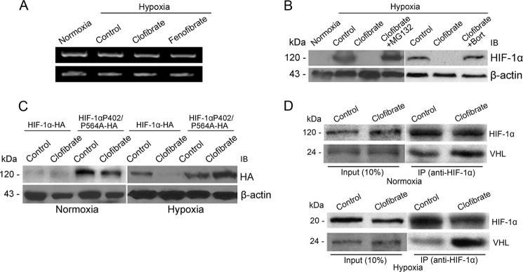
FIGURE 4.
Clofibrate promotes HIF-1α proteasomal degradation. A, MCF-7 cells were treated with 500 μm clofibrate and 100 μm fenofibrate for 4 h prior to placement in a hypoxia chamber for 16 h. RT-PCR was performed to determine the mRNA levels of HIF-1α and β-actin. Shown are representative images of three individual experiments. B, MCF-7 cells were treated with 500 μm clofibrate in the presence or absence of 10 μm MG132 or 1 μm bortezomib (Bort) for 4 h prior to placement in a hypoxia chamber for 16 h. Cell lysates were prepared, and Western blotting was performed using antibodies against HIF-1α and β-actin. Shown are representative images of three individual experiments. IB, immunoblot. C, MCF-7 cells were transfected with either HA-HIF-1α expression vector or HA-HIF-1α(P402A/P564A) mutant vector. After 48 h of transfection, cells were treated with 500 μm clofibrate for 4 h prior to placement in a hypoxia chamber or kept under normoxic conditions for 16 h. Cell lysates were prepared, and Western blotting was performed using antibodies against HA and β-actin. Shown are representative images of three individual experiments. D, MCF-7 cells were treated with 500 μm clofibrate for 4 h prior to addition of 10 μm MG132. The cells were then either placed into a hypoxia chamber or kept under normoxic conditions for 16 h. Cell extracts were prepared, and equal amounts of cell extracts from each sample were immunoprecipitated (IP) with HIF-1α antibody. The immunoprecipitates were separated on a 10% SDS-polyacrylamide gel and blotted with antibodies against HIF-1α and pVHL. Shown are representative images of three individual experiments.

