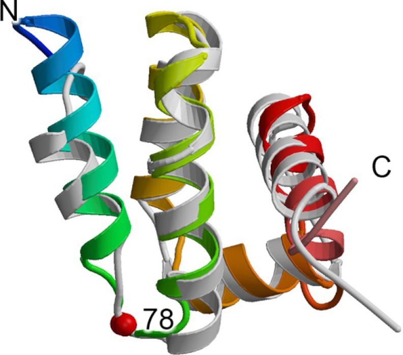
FIGURE 4.
Structure of Sdh5/SdhE. The structure of the yeast Sdh5 fragment (Protein Data Bank code 2LM4)4 is colored with the N terminus in blue and the C terminus in red and is shown overlaid with the structure of E. coli SdhE (Protein Data Bank code 1X6I (81)) colored in light gray. The location of the G78R mutation is highlighted as a red sphere.

