FIGURE 3.
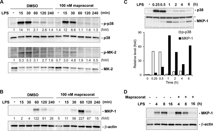
Mapracorat enhances LPS-induced MKP-1 expression and accelerates p38/MK-2 deactivation in Raw 264. 7 cells. A, effects of mapracorat on the kinetics of p38 and MK-2 activation in macrophages following LPS stimulation. Cells were pretreated with 100 nm mapracorat or vehicle (DMSO) for 4 h prior to stimulation with LPS (100 ng/ml). Cell lysates were collected at the indicated time points, and p38 and MK-2 activation was analyzed by Western blot, using antibodies against phospho-p38 (top panel) and phospho-MK-2 (third panel). The membranes were then stripped and blotted with antibodies against total p38 (second panel) or total MK-2 (bottom panel), to verify comparable sample loading. B, effect of mapracorat on the accumulation of MKP-1 shortly after LPS stimulation. Cells were treated as in A, MKP-1 accumulation were examined by Western blot, using an antibody against MKP-1 (upper panel). The membrane was stripped and reprobed with the β-actin antibody (lower panel), for confirming comparable sample loading. In A and B, the intensity of the bands was quantified by densitometry. The signals of the bands of interest were normalized to the loading reference bands. Values presented below the blots represent fold-increase relative to the controls (DMSO treated, without LPS treatment, whose level was set equal to 1). C, dampened oscillatory characteristics of phospho-p38 and MKP-1 levels in macrophages following LPS stimulation. Cells were stimulated with LPS (100 ng/ml) and harvested at the indicated time. Phospho-p38 and MKP-1 were detected by Western blot. Comparable loading is indicated by the similar intensities of the nonspecific band on the MKP-1 blot (marked by an asterisk, lower panel). The intensities of phospho-p38 and MKP-1 were quantified by densitometry. The reciprocal relationship between phospho-p38 and MKP-1 is depicted in the graph underneath the blots. D, effect of mapracorat on MKP-1 accumulation over a longer time frame. LPS (100 ng/ml) was added to the medium together with either vehicle (DMSO) or mapracorat (100 nm) in the cell culture. Cell lysates were prepared at the indicated time points. MKP-1 levels in the lysates were detected by Western blotting. Results presented are representative images.

