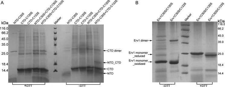
FIGURE 2.
Electrophoresis of the complexes between NTD and CTD mutants. A, the Coomassie-stained gel shows the formation of intermolecular disulfide bonds between NTD-C30S and CTD mutants after incubation under nonreducing conditions (see “Experimental Procedures”). First through fifth lanes, NTD-C30S, CTD-C130S, CTD-C133S, NTD-C30S + CTD-C130S, and NTD-C30S + CTD-C133S, with 5 mm DTT; sixth lane, protein marker; seventh through eleventh lanes, samples corresponding to first through fifth lanes, respectively, without DTT. B, the Coomassie-stained gel shows the formation of mixed disulfide dimer after incubation under nonreducing conditions. First and second lanes, Erv1C30S/C130S and Erv1C30S/C133S, without DTT; third lane, protein marker; fourth and fifth lanes, samples corresponding to first and second lanes, respectively, with 5 mm DTT.

