
FIGURE 4.
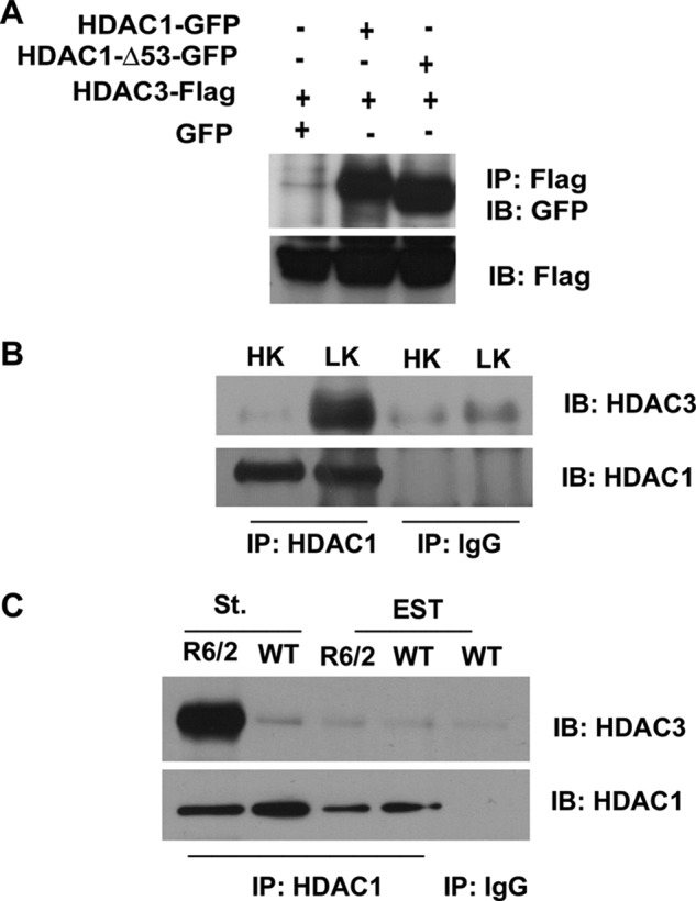
Full-length HDAC1 and HDAC1-Δ53 interact with HDAC3. A, HEK293 cells were co-transfected with FLAG-tagged HDAC3 and GFP, HDAC1, or HDAC1-Δ53 for 24 h, after which lysates were collected and immunoprecipitation (IP) was performed using FLAG antibody. Lysates were subjected to Western blot (IB) analysis and probed with GFP antibody. The same membrane was then reprobed with FLAG antibody to show the immunoprecipitated HDAC3 protein. B, CGNs were treated with either HK or LK for 6 h. Lysates were collected, and immunoprecipitation was performed using either HDAC1 or IgG antibody as negative control. The immunoprecipitate was subjected to Western blot analysis and probed with HDAC3 antibody. The membrane was reprobed with HDAC1 antibody to show pulldown of HDAC1. C, lysates from the striatum (St.) and extra-striatal tissue (EST) of R6/2 mice or their wild-type (WT) littermates were used to immunoprecipitate HDAC1 and were then run on a Western gel and probed with HDAC3 antibody. The membrane was reprobed with HDAC1 to show pulldown of the endogenous protein.