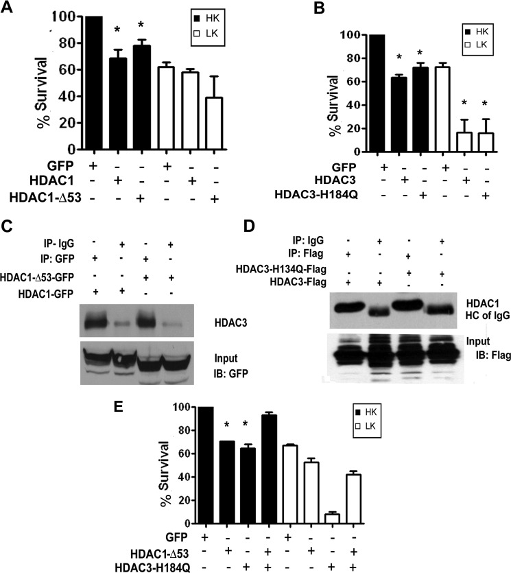
FIGURE 6.
Deacetylase activity of either HDAC1 or HDAC3 Is sufficient for neurotoxicity. A, CGNs transfected with GFP, HDAC1, or HDAC1-Δ53 were treated with HK or LK medium after 8 h. Viability was assessed 24 h later. B, CGNs transfected with GFP, HDAC3, or HDAC3-H134Q were treated with HK or LK medium 8 h later for 24 h, and viability was assessed. C, HEK293 cells were transfected with either GFP-tagged HDAC1 or HDAC1-Δ53. The proteins were immunoprecipitated (IP) using either GFP or IgG antibody and analyzed by Western blotting (IB) with HDAC3 antibody. Both HDAC1 and HDAC1-Δ53 pull down endogenous HDAC3. The lower panel shows the input probed with GFP antibody showing overexpressed HDAC1 proteins. D, lysates from HEK293 cells transfected with FLAG-tagged HDAC3 or HDAC3-H134Q were immunoprecipitated with either FLAG or IgG antibody and analyzed by Western blotting with HDAC1 antibody. Both HDAC3 and HDAC3-H134Q pull down HDAC1. The lower panel shows the input probed with FLAG antibody showing overexpressed HDAC3 proteins. E, CGNs were transfected with GFP, HDAC1-Δ53, or HDAC3-H134Q or co-transfected with HDAC1-Δ53 and HDAC3-H134Q and treated with HK or LK medium after 8 h for 24 h, after which viability was quantified. Error bars in panels A, B, and E indicate S.D.

