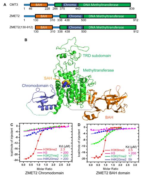
Figure 4. Binding to Methylated Peptides and the Structure of ZMET2 in the Free State.
(A) Color-coded domain architecture of full length CMT3, ZMET2, and the ZMET2 (130-912) construct used to grow crystals.
(B) Ribbon representation of the structure of ZMET2 with bound SAH. The BAH, methyltransferase, and chromo domains are colored in orange, green, and blue, respectively, with the bound SAH molecule shown in a space filling representation. Some disordered regions were not built in the final model and are shown as dashed lines.
(C, D) ITC binding curves for complex formation between ZMET2 chromo (C) and BAH (D) domains and H3K9me2, H3K27me2, H4K20me2, and unmodified H3 peptides. Kd values are listed as an insert. See also Figure S2 and Table S5.

