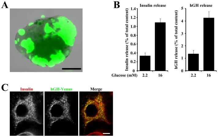
Figure 4. Development of new transfection system.
(A) Representative islets transfected with EGFP. Bar = 100 µm. (B) Pancreatic islets transfected with hGH were stimulated with 2.2 or 16 mM glucose, and the amounts of secreted insulin (left) and hGH (right) from the same batch of islets are expressed as a percentage of the total cellular content. Results are means ± S.E.M. (n = 8). (C) Pancreatic islets were transfected with hGH-venus followed by dispersion on coverslips. Two days after transfection, β-cells expressing hGH-venus were immunostained for insulin. Bar = 5 µm.

