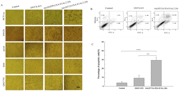
Figure 3. Morphological changes and apoptosis detected by flow cytometry. A.
Morphological observations of tumor cells and normal cells infected with the various oncolytic adenoviruses as detected by microscopy. Cells were infected at an MOI of 10, and the morphological changes in the cells were observed by microscopy after 72 hours of infection. B. Detection of apoptosis in SW620 cells by FACS. SW620 cells were infected with either ONYX-015, Ad·(EGFP)·CEA·E1A(Δ24) or Ad·(ST13)·CEA·E1A(Δ24) at an MOI of 10. At 48 hours, the cells were harvested and stained with annexin V-FITC (for early-stage apoptosis) or PI (for late-stage apoptosis) and were examined by flow cytometry. C. The percentage of apoptotic cells was calculated using the Cell Quest software. The data are presented as the mean ± SD (error bars) of triplicate experiments. (**p<0.01; ***p<0.001).

