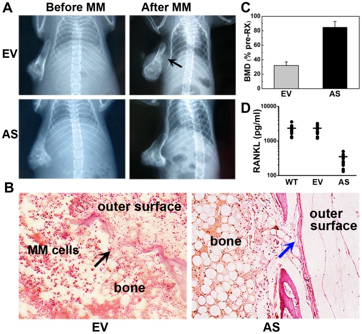
Figure 4. Antisense inhibition of BDNF in ARH77 cells prevents tumor-induced osteolytic lesions in bone implants.
(A) Representative X-ray radiographs of the implanted myelomatous bones in each group, before cell engraftment (Before MM) and at the end of the experiment (After MM). Bones engrafted with EV-ARH cells were severely damaged, and tumors grew on the outer surface of the implanted bone (black arrow). However, no obvious osteolytic destruction was detected in bones harboring AS-ARH cells. (B) H&E staining of bone sections. Bones in the EV-ARH group were severely resorbed. Tumors damaged normal bone structures and infiltrated to the outer surface of the implanted bones (black arrow). In contrast, bones harboring AS-ARH cells had relatively normal structures and continuous cortexes (blue arrow). Magnification ×100. (C) Changes in the BMD of implanted bones. Error bars represent SEMs, P<0.01. (D) Changes in the level of soluble RANKL in rabbit bone implants. The mean levels of RANKL protein expression in WT-, EV-, and AS- group were 2205.9, 2130.5, and 286.3 pg/ml, respectively. RANKL levels in AS- group were significantly reduced when compared to EV- group (P<0.01).

