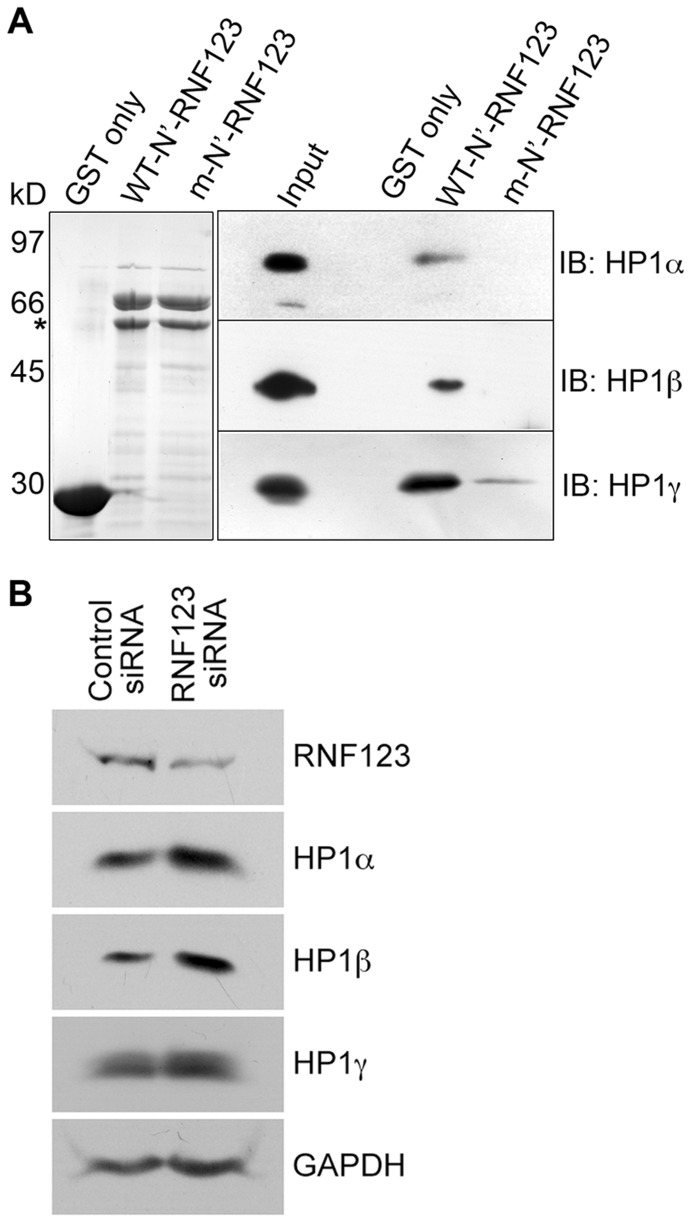
Figure 4. Specificity of RNF123 for HP1 isoforms.
(A) Binding studies with N’-RNF123 and HP1 proteins. Left panel: Coomassie blue-stained gel of purified GST, wild-type and mutant GST-N’-RNF123 proteins. Molecular mass markers: phosphorylase b, 97 kD; albumin, 66 kD; ovalbumin, 45 kD; carbonic anhydrase, 30 kD. The asterisk indicates proteolytic degradation products of GST-N’-RNF123. Right panel: Western blots of bound proteins from HeLa lysates probed with antibodies to HP1 proteins. The input lane represents 1% of total lysate from HeLa cells. (B) Effects of RNF123 siRNA treatment. HeLa were transfected with siRNAs for RNF123 or a control siRNA and lysates were probed with the indicated antibodies by western blot analysis.

