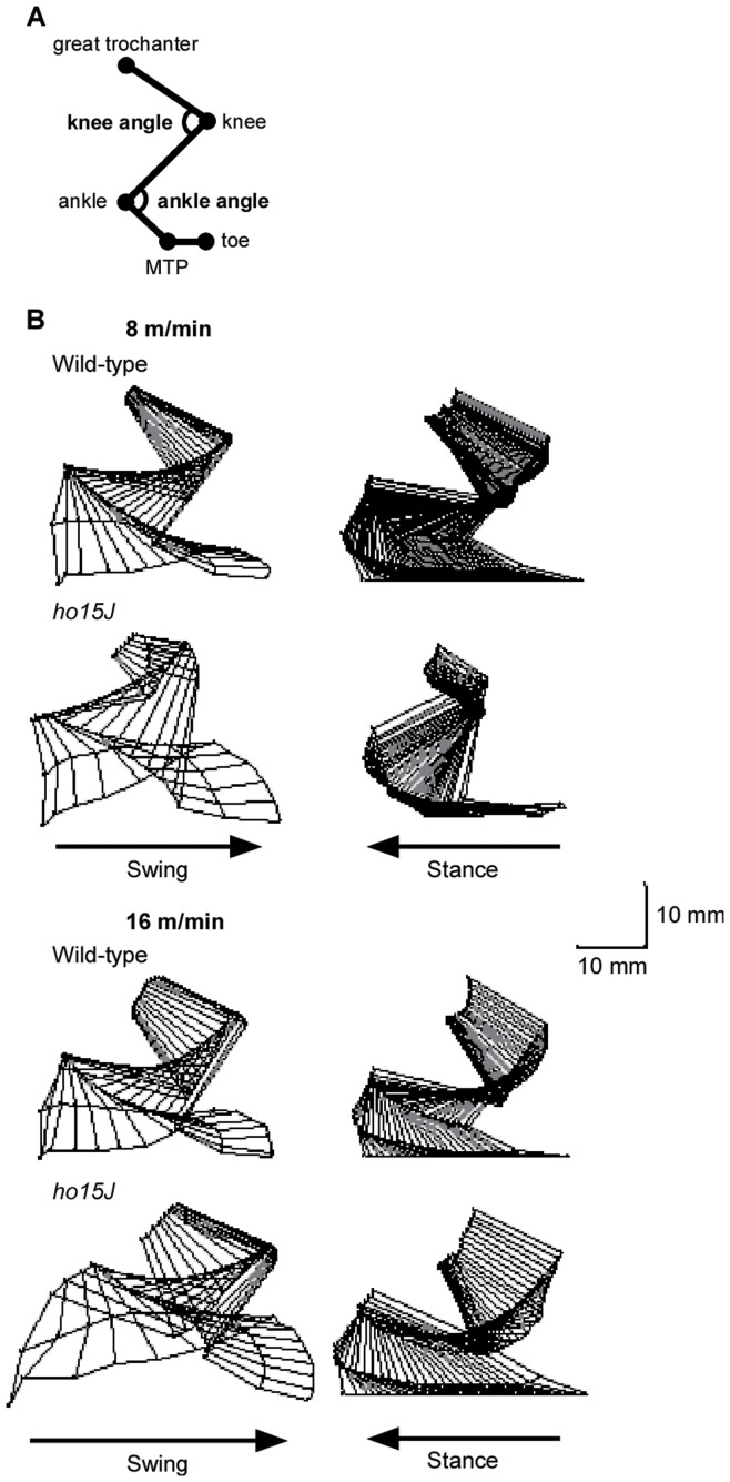
Figure 1. Stick figures of the right hindlimb during the step cycle.
A: Positions of the reflective markers. Reflective markers were placed on the following anatomical landmarks: great trochanter, knee, ankle, 5th metatarsophalangeal joint (MTP), and a toe. B: Typical examples of stick figures of a step cycle (swing and stance phases) at different treadmill speeds for a ho15J mouse and a wild-type mouse. Scale bar indicates 10 mm.

