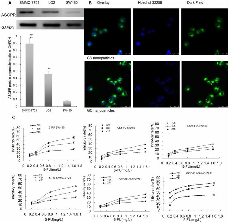
Figure 3. 5-FU nanoparticle enhanced 5-FU to inhibit the growth of hepatic caner through ASGPR -mediated endocytosis in vitro.
(A) ASGPR protein expression was detected by western blot in SMMC-7721, SW480 and LO2 cell lines (n = 3). The ASGPR protein expression in SMMC-7721 and LO2 was stronger than with SW480 (P<0.01), the expression in SMMC-7721 increased significantly than those with SW480 (P<0.01). **P<0.01 compared to SW480, ##P<0.01; compared to LO2. (B) Concurrent focal images of SMMC-7721 cells after 4 h incubation with CS and GC nanoparticles. The strong green fluorescent bright spots in SMMC-7721 cells were observed in the GC nanoparticles using a laser scanning confocal microscope and only small amount of green fluorescent spots were found in the CS nanoparticles without galactosyl ligand modified chitosan. So it was confirmed that the GC nanoparticles targeted liver cancer cells and entered into cells via ASGPR-mediated endocytosis. (C) Comparison of the inhibition rates of 5-FU, CS/5-FU and GC/5-FU on SW480 and SMMC-7721 cells. Results are shown as a average ± means and standard deviation (n = 3). At the 24, 48 and 72 h time points, the rate of tumor inhibition rate decreased from in the order GC/5-FU-SMMC-7721 to 5-FU-SW480 to 5-FU-SMMC-7721 to CS/5-FU-SMMC-7721 to CS/5-FU-SW480 to GC/5-FU-SW480.

