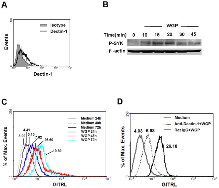
Figure 1. Up-regulation of GITRL on BMDCs via dectin-1 upon WGP stimulation.
(A) Expression of dectin-1 on BMDCs. BMDCs were stained for dectin-1 with anti-dectin-1 antibody (thick line) or rat IgG2b (solid gray) and then analyzed using flow cytometry. (B) Analysis of the activation of SYK by immunoblot of lysates of BMDCs stimulated with WGP (time, above lanes), assessed with anti-phospho-SYK antibody (upper band). β-actin levels were measured as protein loading controls (lower band). (C) Flow cytometry for surface expression of GITRL on BMDCs after no stimulation or stimulation with WGP for 24 h, 48 h and 72 h. (D) BMDCs were pretreated with anti-dectin-1 mAb or rat IgG (5 µg/ml) for 1 h at 37°C and then treated with 100 µg/ml WGP, after 48 h stimulation, cells were subjected to analyze the GITRL expression. The values shown in the flow cytometry profiles are geometric mean fluorescent intensity. Data are representative of three independent experiments.

