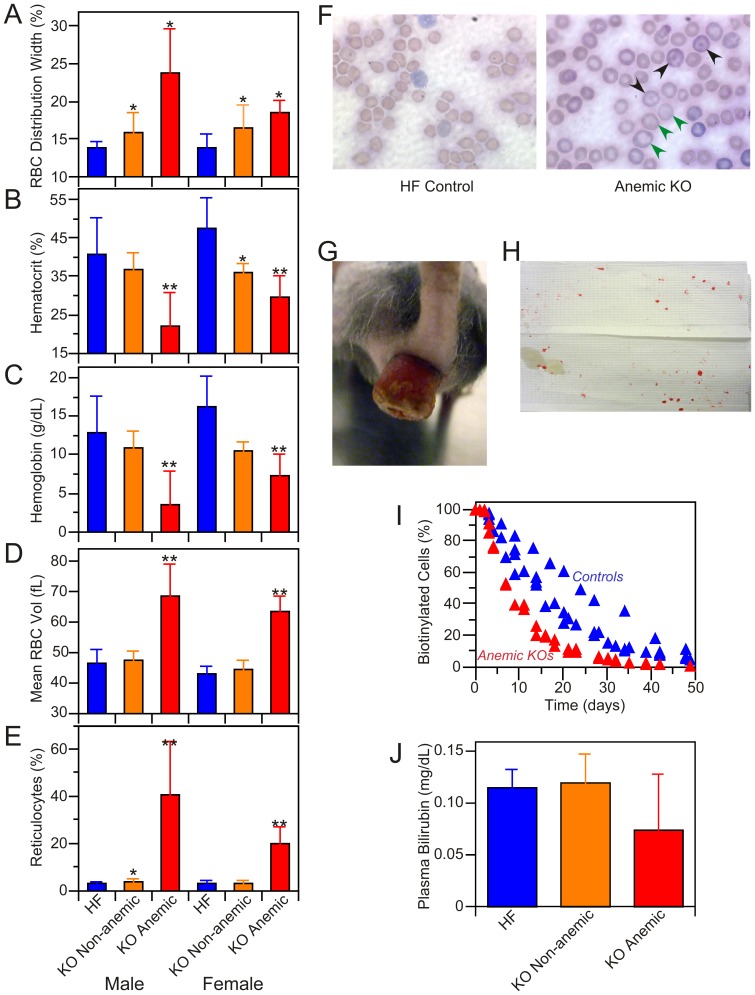
Figure 4. Characterization of anemic KO mice.
Approximately 20% of the KO mice were found to be anemic. A–E: blood cell analysis. A: Red blood cell distribution width. Anemic KO mice (5 males and 5 females) were distinguished from non-anemic KO (20 males and 11 females) and HF control (10 males, 9 females) on the basis of lower hematocrit (panel B), lower hemoglobin level (panel C), enlarged red cells (panel D) and elevated reticulocyte levels (panel E). **Significantly different (p<0.05) from non-anemic KO and HF control mice; *significantly different from HF controls. None of the parameters were significantly different between male and female KOs. No major differences were seen in other blood cell components. The complete blood count analysis is presented as Table S1. F: Wright's-stained blood smears from anemic KO and HF control male mice. Green arrows indicate enlarged erythrocytes, black arrows codocytes. G: Rectal prolapse in KO female, 9 months post-induction; 9 of the 10 anemic mice exhibited rectal prolapse. H. Blood spots on cage floor blot (∼9″×6″) obtained by 5 min exposure to 3 anemic mice with rectal prolapse; no blood spots were observed on blots from cages housing non-anemic KO or HF mice. I: Red cell turnover study performed on 2 anemic KOs and 4 HF control female mice. The two anemic mice had reticulocyte levels of 13.4 and 20.2%. J: Plasma bilirubin levels in HF (n = 6), non-anemic KO (n = 5) and anemic KO (n = 2) mice; differences between groups were not statistically significant.

