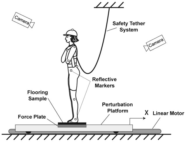
Fig. 1.
Schematic diagram of the experimental setup for the backwards floor translation task. A force plate measured the time-varying location of the underfoot centre of pressure, while a motion capture system measured the position of 23 markers attached to the following anatomical landmarks: top of the head, spinous process of the C7 vertebra, sacrum, bilaterally on the acromion, lateral epicondyle of the humerus, styloid process of the radius, anterior superior iliac spine, greater trochanter, lateral condyle of the femur, calcaneus, lateral malleolus, head of the fifth metatarsal, and distal phalange of hallux.

