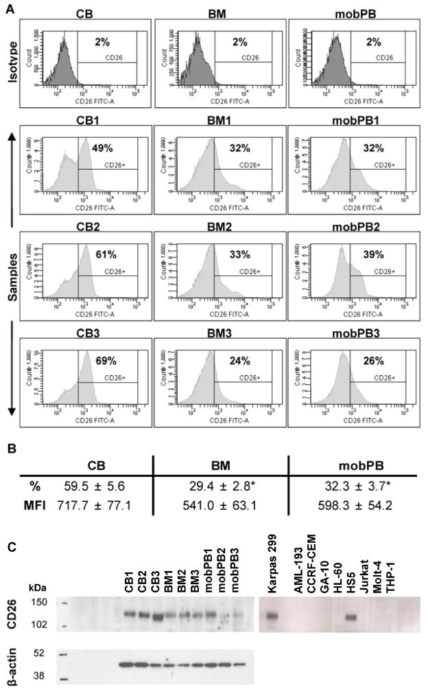
Figure 2. CD26 expression on non-adherent MNC by flow cytometry and Western blot analysis.
(A) Flow cytometry was performed on non-adherent CB, BM, and G-CSF mobPB MNC using fluorochrome-conjugated monoclonal antibodies. Representative isotype and sample cell count vs. CD26-FITC histograms are shown. (B) Average CD26 expression and MFI are shown. *P≤0.05 as compared to CB MNC. (C) CD26 expression in corresponding non-adherent MNC and cell lines by Western blot. The representative molecular weight markers (kDa) are shown to the left of each blot. When probed with anti-hCD26/DPPIV goat polyclonal IgG, CD26 (110 kDa) bands appeared at various intensities for CB, BM, and G-CSF mobPB MNC. In the cell lines, Karpas 299 was positive for CD26 and Jurkat was negative, as previously reported. In addition, HS5 cell line was positive for CD26, and all others tested were negative. Both blots were re-probed with anti-β-actin (42 kDa) to ensure equal loading.

