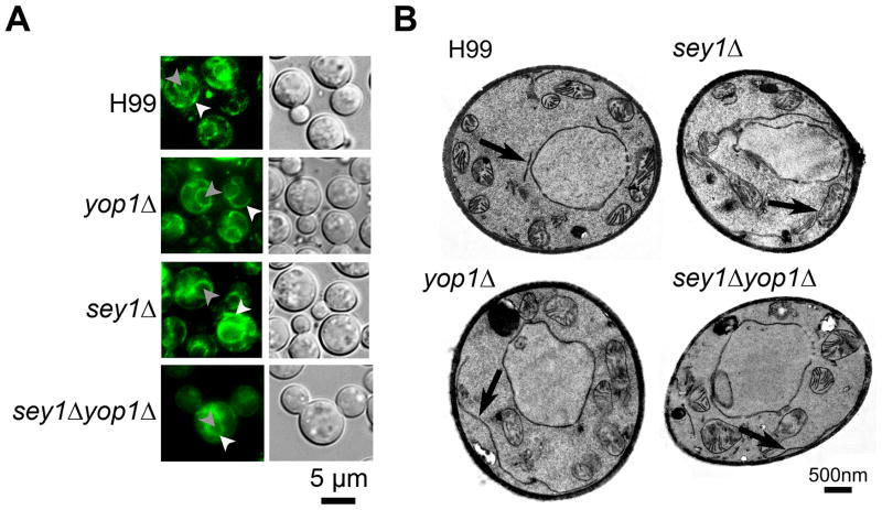
Figure 1. Disruption of YOP1 causes ER abnormality as was seen in sey1Δ.
A) Localization of the ER protein GFP-Sec61β in each indicated strain. Cells were grown on YPD agar at 30°C and subsequently visualized by fluorescence microscopy. White arrow head = ER; grey arrow head = nucleus (determined by co-localization with Hoechst 3342 dye as published previously (Ngamskulrungroj, et al., 2012)). B) Transmission Electron Microscopy (TEM). Overnight cell cultures of indicated strains grown to mid log phase were prepared and examined by TEM. Arrow = ER.

