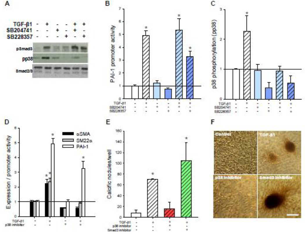
Fig. 2. 5-HT2B antagonism prevents non-canonical p38 MAPK but not canonical Smad3 signaling.
A, TGF-β1 treatment leads to increased Smad3 phosphorylation (pSmad3) after 15 min and p38 phosphorylation (pp38) after 1 h. 5-HT2B antagonism inhibits TGF-β1-induced pp38 but not pSmad3. B, 24 h of TGF-β1 treatment leads to a five-fold increase in canonical PAI-1 promoter activity that is not inhibited by either of the 5-HT2B antagonists (n ≥ 3). C, Average densitometry (n = 5) reveals a two-fold increase in pp38 after TGF-β1 treatment that is completely inhibited by 5-HT2B antagonism. D, p38 inhibitor blocks TGF-β1-induced αSMA expression and SM22α promoter activity but not PAI-1 promoter activity. e, p38 inhibition blocks TGF-β1-induced calcific nodule morphogenesis but Smad3 inhibitor does not. d, Representative images indicate the presence of calcific nodules in AVICs treated with TGF-β1 alone or in combination with Smad3 or p38 inhibitors. All error bars indicate standard error of the mean. * indicates significant difference (p < 0.005) versus control. Scale bar = 250 µm.

