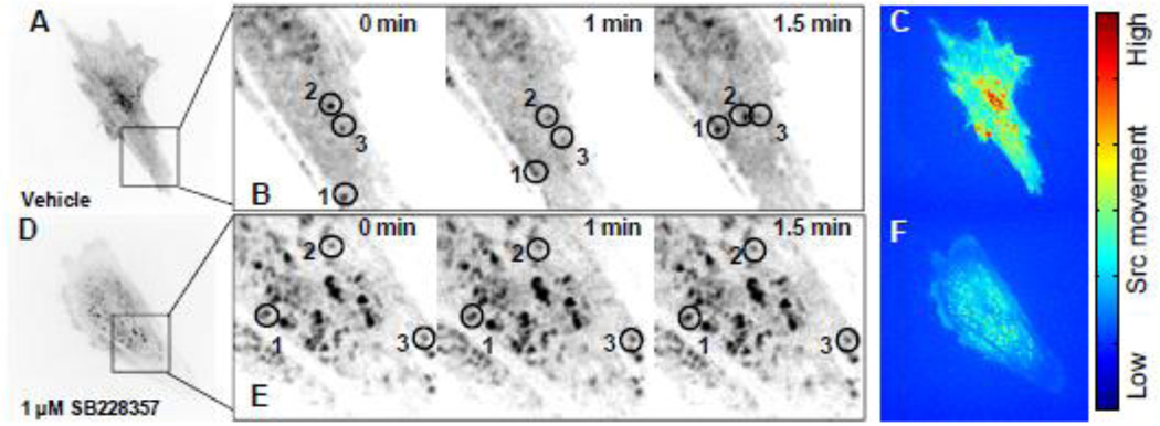
Fig. 4. 5-HT2B antagonism arrests Src motility within AVICs.
A, AVICs transfected with mCherry-N1-Src and treated with DMSO vehicle for 1 h were imaged by time-lapse microscopy. Images were taken every 15 s for a total of 15 min. B, A subsection of a DMSO vehicle treated AVIC allows for clear identification of Src movement at t = 0, 1, and 1.5 min. C, Eulerian based analysis of pixel intensity changes within the image over the course of 15 min represents Src kinematics. Red indicates a high degree of movement, and blue indicates little movement within a pixel. D, E, AVICs treated with 1 µM SB228357 for 1 h exhibit little to no Src movement at t = 0, 1, and 1.5 min. F, 5-HT2B antagonism arrests Src movement over the 15 min imaging time.

